| |
|
|
|
|
|
|
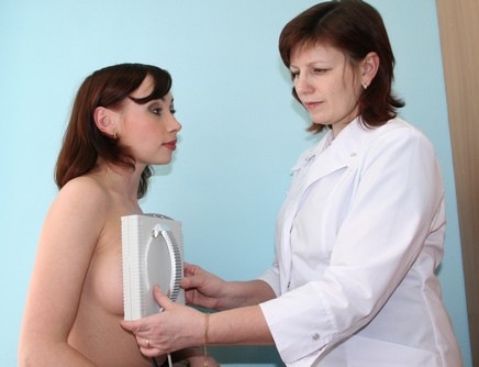
Carrying out measurement |
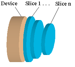
Realization of the 3D imaging |
|
|
|
|
|
EIT image of the normal breast, the slice is on 1.2 cm depth, the nipple is in the center |
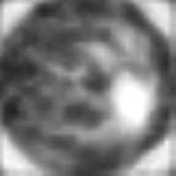
EIT image of the breast with large carcinoma, the slice is on 1.2 cm depth |
Information about Multifrequency Electrical impedance Mammograph MEM
|
|
||
| |
|
|